الشرح
Anatomical Pathway and Structures of the Respiratory Tract
The human respiratory system is composed of a specialized network of passages and organs designed to move atmospheric air deep into the body where oxygen can be transferred to the bloodstream and carbon dioxide can be removed. Air enters the body primarily through the nasal cavity or the oral cavity, where it is warmed, moistened, and filtered by tiny hairs called vibrissae and sticky mucus. The air then travels down into the pharynx, which serves as a shared pathway for both food and air, before passing through the larynx, commonly known as the voice box, which contains the vocal cords and is protected by a flap of tissue called the epiglottis that prevents food from entering the airway during swallowing.
From the larynx, air moves into the trachea, a strong, flexible tube reinforced by rings of cartilage in the shape of the letter C. These incomplete cartilaginous rings prevent the trachea from collapsing during pressure changes while allowing the adjacent oesophagus to expand when food is swallowed. The inner lining of the trachea is coated with ciliated epithelial cells and mucus-producing goblet cells. The mucus traps dust, bacteria, and other foreign particles, while the beating cilia continuously sweep this contaminated mucus upward toward the throat, where it is swallowed and destroyed by stomach acid.
At the lower end of the trachea, the airway branches into two primary bronchi, known individually as a bronchus, with one bronchus entering each lung. Within the lungs, these primary bronchi subdivide repeatedly into increasingly smaller tubes called bronchioles. Unlike the trachea and primary bronchi, the smallest bronchioles lack cartilage support and instead possess smooth muscle in their walls, which can contract or relax to regulate the flow of air into the respiratory zones.
Lung Structure and Alveolar Specialisations for Gas Exchange
The lungs are spongy, elastic organs enclosed within the thoracic cavity, protected by the ribcage and separated from the abdomen by the dome-shaped diaphragm muscle. The right lung is divided into three lobes, while the slightly smaller left lung has two lobes to accommodate the position of the heart. Each lung is surrounded by a double-layered membrane called the pleural membranes, enclosing a fluid-filled pleural cavity that reduces friction as the lungs expand and contract during ventilation.
Deep within the lung tissue, the terminal bronchioles terminate in clusters of microscopic air sacs known as alveoli. The millions of alveoli present in human lungs provide an enormous combined surface area for the exchange of gases between the air and the circulatory system. The structure of the alveoli is exceptionally well adapted for efficient gas exchange through several key features. First, the walls of the alveoli are extremely thin, consisting of a single layer of flattened epithelial cells known as squamous epithelium. Second, each alveolus is surrounded by an extensive, dense network of tiny blood vessels called pulmonary capillaries, which also possess walls only one cell thick, creating a very short diffusion distance between the alveolar air and the deoxygenated blood.
Furthermore, the inner lining of each alveolus is covered by a thin film of moisture. Oxygen gas from inhaled air dissolves in this aqueous layer before it can diffuse across the alveolar and capillary walls into the bloodstream. Finally, the continuous flow of blood through the capillary network maintains a steep concentration gradient, ensuring that oxygen is constantly carried away from the exchange surface while carbon dioxide is continuously brought toward it.
The Mechanics of Ventilation: Breathing In and Out
Ventilation, commonly referred to as breathing, is the mechanical process by which air is moved into and out of the lungs to maintain a fresh supply of oxygen and remove carbon dioxide. This process is driven by changes in volume and pressure within the thoracic cavity, achieved through the coordinated contraction and relaxation of two primary muscle groups: the diaphragm and the intercostal muscles located between the ribs.
During inhalation, or inspiration, an active muscular contraction takes place. The dome-shaped diaphragm contracts and flattens downward, increasing the vertical dimension of the thoracic cavity. Simultaneously, the external intercostal muscles contract, pulling the ribcage upward and outward. These combined movements increase the overall volume of the thorax. According to Boyle's law, as the volume of a closed container increases, the pressure of the gas inside decreases. Therefore, the air pressure inside the lungs drops below atmospheric pressure. This pressure gradient forces atmospheric air to rush down the trachea and bronchi into the lungs until the internal and external pressures equalize.
During exhalation, or expiration under resting conditions, the process is largely passive. The diaphragm relaxes and returns to its dome-shaped upward position, while the external intercostal muscles relax, allowing the ribcage to move downward and inward under the influence of gravity and the elastic recoil of the lung tissue. This decreases the volume of the thoracic cavity, which in turn increases the internal air pressure above atmospheric pressure. The higher pressure inside the lungs forces air outward through the respiratory tract into the atmosphere. During forced exhalation, such as during vigorous physical exercise, internal intercostal muscles and abdominal wall muscles contract actively to push air out more rapidly.
Gas Exchange via Diffusion Across the Respiratory Membrane
Gas exchange is the physical process by which oxygen enters the bloodstream and carbon dioxide is removed from it, occurring across the ultra-thin respiratory membrane formed by the alveolar and capillary walls. This entire exchange relies exclusively on the process of diffusion, which is the passive net movement of particles from a region of higher concentration to a region of lower concentration down a concentration gradient, requiring no metabolic energy input from the organism.
Inhaled air in the alveoli has a high concentration and partial pressure of oxygen and a low concentration of carbon dioxide. Conversely, the deoxygenated blood arriving at the pulmonary capillaries from the right side of the heart has travelled from actively respiring body tissues, resulting in a low concentration of oxygen and a high concentration of carbon dioxide. Because of these concentration differences, oxygen molecules dissolve in the moist alveolar lining, diffuse across the single-celled alveolar epithelium and capillary endothelium, and bind to haemoglobin inside red blood cells.
Simultaneously, carbon dioxide dissolved in the blood plasma and carried as bicarbonate ions moves in the opposite direction. Because the partial pressure of carbon dioxide is higher in the incoming capillary blood than in the alveolar air, carbon dioxide diffuses out of the blood plasma, crosses the capillary and alveolar walls, enters the alveolar air space, and is subsequently expelled from the body during exhalation. This dual directional diffusion operates continuously and efficiently due to the massive surface area, short diffusion distance, and maintained concentration gradients provided by uninterrupted pulmonary ventilation and blood circulation.
Distinction Between Breathing and Respiration
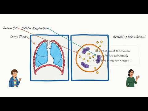
It is scientifically crucial to distinguish between breathing and cellular respiration, as everyday language often uses the two terms interchangeably. Breathing, or ventilation, is a purely physical and mechanical process involving the muscular movement of the ribcage and diaphragm to ventilate the lungs with atmospheric air. It takes place at the organ level and serves to supply oxygen to the respiratory surfaces and remove waste gases.
Cellular respiration, on the other hand, is a vital biochemical process that occurs at the cellular level inside every living cell, specifically within organelles called mitochondria. During cellular respiration, glucose and oxygen undergo a series of enzyme-controlled chemical reactions to release usable chemical energy in the form of adenosine triphosphate (ATP) to drive cellular activities. The overall chemical reaction produces carbon dioxide and water as metabolic waste products.
While breathing provides the raw oxygen required for aerobic cellular respiration and eliminates the carbon dioxide produced as a waste product, the two processes are distinct in mechanism and location. Breathing is macroscopic, physical, and involves skeletal muscles and pressure gradients, whereas cellular respiration is microscopic, chemical, and takes place within intracellular fluid and mitochondria. Without breathing, the body's cells would quickly be starved of the oxygen needed for respiration, and metabolic poisons would accumulate.